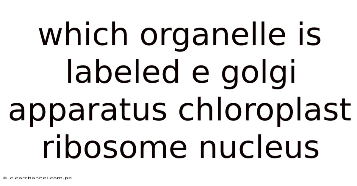
Which Organelle Is Labeled “E”? – Golgi Apparatus, Chloroplast, Ribosome, or Nucleus?
Once you open a typical biology textbook or slide‑presentation on cell structure, you’ll often see a schematic of a eukaryotic cell with letters marking each component. One of the most common questions students encounter is: *“In this diagram, which organelle is labeled ‘E’?Worth adding: * The answer can vary depending on the illustration, but the four most frequent candidates are the Golgi apparatus, chloroplast, ribosome, and nucleus. Understanding how to identify each of these organelles—and why they might be chosen for the “E” label—helps you not only ace exams but also appreciate the functional diversity inside a cell.
Below we break down the distinguishing features of each organelle, discuss the contexts in which “E” typically points to one of them, and provide a quick decision‑tree you can use during tests or lab work. By the end of this article you’ll be able to look at any cell diagram and instantly know which structure the mysterious “E” represents Easy to understand, harder to ignore..
1. The Golgi Apparatus – The Cell’s Shipping Center
1.1 What It Looks Like in a Diagram
- Shape: Stacked, flattened membrane sacs (cisternae) that often appear as a curved ribbon or a series of pancake‑like layers.
- Location: Usually situated near the nucleus, between the endoplasmic reticulum (ER) and the plasma membrane.
- Labeling convention: Many textbooks label the Golgi with letters E, F, or G because it sits in the “central” region of the cell diagram.
1.2 Key Functions
- Protein modification: Adds carbohydrate groups (glycosylation) and other chemical tags to proteins received from the rough ER.
- Sorting and packaging: Packages modified proteins into vesicles destined for secretion, lysosomes, or the plasma membrane.
- Lipid transport: Modifies and distributes lipids, especially in plant cells where it helps form the cuticle.
1.3 When “E” Is Likely the Golgi
- The diagram shows a curved stack of membranes rather than a solid sphere.
- The organelle is adjacent to the nucleus but not inside it.
- There are arrows indicating vesicle traffic moving away from this structure, a hallmark of Golgi activity.
2. Chloroplast – The Green Powerhouse of Plant Cells
2.1 Visual Cues in Illustrations
- Shape: Oval or disc‑shaped, often larger than other organelles, with a double membrane.
- Internal structure: Visible thylakoid stacks (grana) and a fluid stroma; sometimes drawn as green shading to hint at chlorophyll.
- Label placement: In plant‑cell diagrams, letters E or C frequently point to the chloroplast because it is the most distinctive organelle.
2.2 Core Roles
- Photosynthesis: Captures light energy to convert CO₂ and H₂O into glucose and O₂.
- Synthesis of fatty acids and amino acids: Provides building blocks for the cell’s metabolism.
- Storage: Accumulates starch granules during periods of excess photosynthate.
2.3 When “E” Means Chloroplast
- The cell is plant or algal, not animal.
- The labeled structure is green‑tinted and contains internal stacks (grana).
- The diagram may include light rays or CO₂ arrows pointing toward the organelle, emphasizing its photosynthetic role.
3. Ribosome – The Molecular Factory for Protein Synthesis
3.1 How Ribosomes Appear in Cell Sketches
- Size: Very small, often depicted as tiny dots or spheres.
- Distribution: Either free in the cytoplasm (scattered dots) or bound to the rough ER (small dots on the ER surface).
- Labeling tendency: Because they are numerous, textbooks rarely assign a single letter to a ribosome; however, in simplified diagrams they may label a cluster of dots as “E” to illustrate translation.
3.2 Functional Highlights
- Protein assembly: Reads mRNA sequences and links amino acids together to form polypeptide chains.
- Two types:
- Free ribosomes produce proteins that function in the cytosol.
- Bound ribosomes (on rough ER) synthesize secretory and membrane proteins.
- Rapid turnover: Ribosomes are assembled in the nucleolus and can be recycled quickly.
3.3 When “E” Refers to Ribosomes
- The diagram shows a cluster of tiny circles either floating in the cytoplasm or attached to a membrane.
- There may be arrows from DNA → mRNA → ribosome, highlighting the central dogma.
- The cell is often bacterial or prokaryotic, where ribosomes are the most prominent organelle.
4. Nucleus – The Command Center
4.1 Recognizing the Nucleus in Images
- Shape: Large, roughly spherical or oval structure, often the biggest organelle in the cell.
- Membrane: Surrounded by a double nuclear envelope with visible pores.
- Internal feature: May contain a nucleolus (a dense, darker spot).
- Label usage: Many textbooks label the nucleus with A, B, or C, but some diagrams—especially those focusing on sub‑nuclear structures—use E for the nucleolus or a specific nuclear region.
4.2 Principal Functions
- Genetic storage: Houses DNA organized into chromosomes.
- Transcription hub: Synthesizes precursor mRNA (pre‑mRNA) that will be processed and exported.
- Regulation: Controls cell cycle, growth, and response to external signals through gene expression.
4.3 When “E” Points to the Nucleus
- The labeled organelle is the largest, centrally located structure with a clear double membrane.
- There may be arrows indicating DNA → RNA or RNA export pathways.
- In a diagram emphasizing nuclear sub‑structures, “E” could specifically denote the nucleolus where ribosomal RNA is assembled.
5. Decision‑Tree: Quickly Identify “E”
-
Is the cell a plant/algal cell?
- Yes → Look for a green, oval organelle with internal stacks → Chloroplast.
- No → Proceed to step 2.
-
Is the labeled structure a large, double‑membrane sphere?
- Yes → It’s the Nucleus (or nucleolus if the image highlights a dense core).
- No → Continue.
-
Does the organelle appear as a curved stack of flattened sacs?
- Yes → That’s the Golgi apparatus.
- No → Move on.
-
Are you seeing tiny dots, either free or attached to a membrane?
- Yes → Those are Ribosomes (free or rough‑ER bound).
- No → Re‑examine the diagram; sometimes “E” may label a less common structure (e.g., lysosome, peroxisome) depending on the textbook.
6. Frequently Asked Questions
6.1 Why do textbooks use different letters for the same organelle?
Authors aim to avoid clutter and to highlight the organelles most relevant to the chapter’s focus. In a chapter on photosynthesis, the chloroplast will likely receive a prominent label, while a chapter on protein trafficking will highlight the Golgi.
6.2 Can the same diagram label “E” for two different organelles in separate figures?
Absolutely. The letter is merely a placeholder; its meaning is defined by the figure legend. Always read the legend first Easy to understand, harder to ignore..
6.3 How can I remember which organelle is which when studying for exams?
Create a simple mnemonic based on shape and function:
- Golgi – Graduated stacks, Goods shipping.
- Chloroplast – Colorful, Carbon fixation.
- Ribosome – Round dots, Reading RNA.
- Nucleus – Neural‑like control center, Needs DNA.
6.4 Do prokaryotes have any of these organelles?
Prokaryotes lack a true nucleus, Golgi apparatus, and chloroplasts. They do possess ribosomes, but these are smaller (70S) and not membrane‑bound.
6.5 What if the diagram shows a “E” next to a membrane‑bound sac that isn’t stacked?
That could be a vacuole or lysosome. Check the legend; if the organelle is involved in waste degradation, it’s a lysosome, whereas a large central sac in plant cells is a vacuole.
7. Conclusion
Identifying the organelle labeled “E” hinges on recognizing shape, size, color, and cellular context. In most educational diagrams, “E” will correspond to one of four major structures:
- Golgi apparatus – the stacked, ribbon‑like shipping hub near the nucleus.
- Chloroplast – the green, disc‑shaped photosynthetic engine of plant cells.
- Ribosome – the minute dots that synthesize proteins, either free or on rough ER.
- Nucleus – the large, double‑membrane command center containing DNA.
By applying the decision‑tree and mnemonic strategies outlined above, you can confidently decode any cell illustration, whether you’re tackling a high‑school biology test, preparing a university lab report, or simply satisfying your curiosity about the microscopic world. Here's the thing — remember: the key is not memorizing letters, but understanding each organelle’s unique visual signature and role. With this knowledge, the mystery of “E” becomes a stepping stone toward mastering cell biology Worth keeping that in mind..